Introduction to Migraine and Brain Structure
Understanding Migraine
Migraine is a neurological disorder characterized by recurrent, moderate to severe headaches, often accompanied by nausea, vomiting, and extreme sensitivity to light and sound. While the exact causes of migraine remain somewhat elusive, growing evidence suggests a complex interplay of genetic predisposition, environmental factors, and alterations in brain function. Understanding the intricacies of brain structure and its potential role in migraine is crucial for developing effective diagnostic and therapeutic strategies.
The Role of the Trigeminal System
The trigeminal nerve plays a pivotal role in migraine, transmitting pain signals from the head and face to the brain. Research indicates that during a migraine attack, the trigeminal system exhibits heightened activity, potentially leading to the characteristic throbbing pain and other associated symptoms. This heightened activity may be linked to specific regions and structures within the trigeminal pathway, suggesting a potential structural difference in migraineurs compared to healthy individuals.
Cerebral Blood Flow Variations
Changes in cerebral blood flow (CBF) are frequently observed in individuals experiencing migraine. These fluctuations can manifest as both increased and decreased blood flow in various brain regions. Understanding the specific patterns and locations of these CBF variations is important to determine if they correlate with specific migraine subtypes and their associated symptoms. Precise mapping of these changes could provide insights into the underlying mechanisms driving migraine pain.
The Amygdala's Potential Involvement
The amygdala, a brain region crucial for processing emotions, particularly fear and anxiety, is increasingly being implicated in migraine pathophysiology. Studies have hinted at a possible connection between heightened amygdala activity and the onset of migraine attacks. This suggests that emotional factors might play a role in triggering migraine episodes, potentially influencing the interplay between brain structure and migraine susceptibility.
The Role of the Cerebellum
The cerebellum, often associated with motor coordination, is also emerging as a potential player in migraine. Some research suggests a possible link between cerebellar dysfunction and the development of migraine. Further investigation is needed to fully elucidate the precise role of the cerebellum in the complex processes leading to migraine attacks and to determine if structural differences exist in the cerebellum of migraineurs.
The Influence of Genetics and Environment
Genetic predisposition is widely recognized as a contributing factor to migraine development. Studies have identified specific genes that might increase the risk of developing migraine. However, environmental factors, such as stress, sleep deprivation, and dietary triggers, also play significant roles in modulating the expression of these genetic predispositions. Understanding the complex interplay between genetics, environment, and brain structure is essential for comprehending the multifaceted nature of migraine.
Neuroimaging Studies and Grey Matter Density
Neuroimaging Techniques
Neuroimaging techniques provide invaluable insights into the structure and function of the brain, offering a window into the complex interplay of neural processes. These techniques, including functional magnetic resonance imaging (fMRI), positron emission tomography (PET), and electroencephalography (EEG), allow researchers to observe brain activity in real-time. This real-time observation is crucial for understanding the dynamic nature of brain function and how it relates to various cognitive processes.
Different neuroimaging methods offer unique advantages. For instance, fMRI provides high spatial resolution, allowing researchers to pinpoint specific brain regions involved in a task. Conversely, EEG offers excellent temporal resolution, capturing rapid changes in brain activity with millisecond precision. This combination of techniques is often used in research to gain a comprehensive understanding of the brain.
Grey Matter Structure
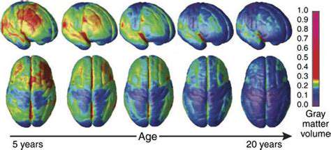
Grey matter, comprised primarily of neuronal cell bodies, dendrites, and synapses, is essential for information processing within the brain. It plays a vital role in various cognitive functions, including learning, memory, and decision-making. Understanding the intricacies of grey matter structure and its relationship to cognitive abilities is a key area of research in neuroscience.
The intricate network of connections within grey matter determines how information is processed and communicated throughout the brain. Variations in grey matter volume or density have been correlated with differences in cognitive performance. This implies a potential link between the structural aspects of grey matter and individual cognitive capabilities.
Neuroimaging and Grey Matter Volume
Neuroimaging studies often investigate the relationship between grey matter volume and cognitive abilities. Researchers examine how variations in the amount of grey matter in specific brain regions correlate with performance on different cognitive tasks. This exploration is crucial for understanding the neural basis of cognitive functions.
Grey Matter and Cognitive Function
Research on grey matter and cognitive function is consistently revealing new insights into the interplay between brain structure and cognitive performance. Studies demonstrate a significant correlation between grey matter volume in specific brain regions and particular cognitive skills. This suggests that the neural circuitry underlying cognitive processes is influenced by the structural characteristics of grey matter.
Further investigation is needed to establish causal relationships between grey matter structure and cognitive function. While correlations are observed, determining if alterations in grey matter structure directly cause changes in cognitive performance remains a complex challenge.
Neuroimaging and Brain Disorders
Neuroimaging techniques play a crucial role in the diagnosis and understanding of various neurological and psychiatric disorders. By visualizing brain structure and function, researchers can identify abnormalities that may contribute to these conditions. This allows for early detection and potentially more effective interventions.
For instance, neuroimaging studies have been instrumental in identifying specific brain regions affected in conditions like Alzheimer's disease or depression. This knowledge is vital for developing targeted therapies and treatment strategies. These advancements highlight the importance of neuroimaging in advancing our understanding and care of brain disorders.